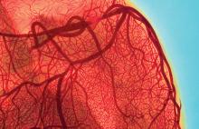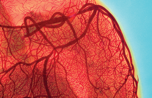
Atherosclerotic plaques don’t form just anywhere; they are more likely to occur at stagnation points in large blood vessels, where the flow of blood is disturbed. Investigating the mechanical principles underlying the response of living cells to fluid flow and its relation to atherosclerosis are the focus of experimental biophysicist Aurelia Honerkamp-Smith.
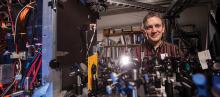
Honerkamp-Smith, assistant professor of physics, combines fluorescence microscopy, lipid physical chemistry and fluid mechanics to study the biophysics of cell membranes. Her lab examines the membrane as a system in its own right to consider flow-mediated membrane protein sorting at the intersection of fluid mechanics and membrane physical chemistry.
“A lot of people think proteins do everything interesting and the membrane is just a substrate for proteins to sit in,” she says. “I want to show that lipids are also important and have interesting properties, and we need to understand how they work, how they are put together, what the energies are of the various interactions.”
Her work crosses scientific disciplines, but physics is the best place for her work because she focuses on the mechanical properties of membrane proteins and lipids, she says. Her work explores the interactions between molecules of different types, the flow of lipids and the forces and energies involved in these interactions. Understanding these interactions is important since proteins and lipids affect how cells function and, thus, our health.
In the lab, she forms vesicles, consisting of fluid enclosed by a lipid bilayer, which she applies to a glass surface. This glass surface is put inside a microscopic channel. She applies a flow and watches then to see how the bilayer responds. By manipulating these interactions, Honerkamp-Smith hopes to determine how much force is needed to force a protein to move. It is also a way to study protein/protein interactions. If proteins are stacked on the downstream edge, eventually they should repel each other. She can then adjust the repulsion properties and, by observing their movement under a fluorescence microscope, measure how much they push each other away.
“When proteins stick out of the membrane, they can act like a sail and move relative to the lipids, as long as there is enough friction keeping the lipids from moving as well,” she says. “This has been observed on the surfaces themselves, but nobody has studied it quantitatively. It hasn’t been studied as a possible mechanism for cell signaling. It’s an interesting physical process that could give us information about what is happening outside the cell. We don’t know if the cells take advantage of this or not.”
Critical forces in lipid membranes are governed not only by membrane properties, but also by how the membrane couples to the surrounding bulk fluid. These basic cellular properties are of interest to biophysicists. The work underway by Honerkamp-Smith and her colleagues will increase our understanding of how cells sense their environment and may lead to improved clinical therapies in the treatment of heart disease.